22/08/2018
Print PageMini guts reveal causes of rare intestinal disease
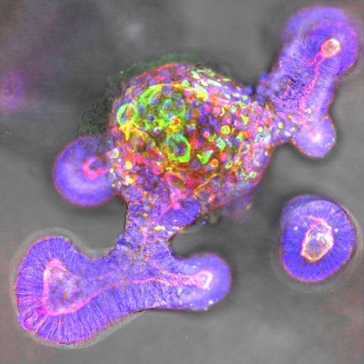
Microvillus Inclusion Disease (MVID) is a congenital disease characterized by chronic diarrhea that prevents the absorption of nutrients via the intestinal villi. In affected individuals, the brush border of microvilli, which increases the internal surface area of the intestinal walls, is either not present at birth, or is lost in the first few weeks of life. This has drastic consequences for newborns and there are only limited treatment options available. Earlier studies have already identified a defect in the cellular transport of microvilli vesicles as an underlying mechanism. These are small, intracellular sacs that are responsible for transporting nutrients in cells. Until now, however, there were no suitable cellular models that could be used to investigate the causes of this defect. The intracellular microvillus inclusions (MVI) that are characteristic of the disease are only rarely observed in cell cultures. As part of an international collaborative project, German Cancer Consortium scientists working at the Georg Speyer Haus in Frankfurt have now used a 3D intestinal organoid culture model to investigate MVID. The researchers discovered that mutations in the MUNC18-2/STXBP2 protein cause different forms of MVID. In organoids – 3D mini guts derived from mouse cells – the scientists were able to demonstrate that the different levels of severity of the disease are linked to the degree of differentiation of the intestinal cells. Using video microscopy, they were able to visualize the process of MVI formation in real time for the first time. This showed an unexpected mechanism of MVI formation in the cytoplasm or through the inclusion of plasma membranes. This culture model could improve the diagnosis of MVID cases in future and be employed to test treatment options using patient-specific organoid models.
Original publication
M.H. Mosa et al., 2018. Dynamic formation of microvillus inclusions during enterocyte differentiation in Munc18-2 deficient intestinal organoids. In: Cellular and Molecular Gastroenterology and Hepatology. Online Publication: 14 August 2018. DOI: https://doi.org/10.1016/j.jcmgh.2018.08.001